and licensed under a Creative Commons Attribution-ShareAlike 4.0 International License.
|
Lesson Three
|
|
Mitosis is specifically designed to distribute chromosomes equally. Mitosis distributes exact
copies of genetic material so the daughter nuclei are genetically
identical to each other and identical to the mother nucleus
from which they came.
The main function of mitosis is to increase the number of identical nuclei. When followed by cytokinesis, as it usually is, mitosis increases
cell numbers. But you know all that - because I told you.
I also told you that meiosis is involved in reproduction but you may have gone away thinking that all organisms must use meiosis to reproduce. That is not true. Many species, including some multicellular species, can reproduce by mitosis and when they do so they produce exact copies of the parents. These are called clones and this method of reproduction is called asexual reproduction. Clones are produced without sex and without meiosis. Humans don't reproduce by cloning (yet) but some animals do, especially invertebrates. Plants are "masters" at cloning, especially with help from gardeners, and that is why asexual reproduction is often called vegetative reproduction or vegetative propagation. All asexual reproduction (cloning, vegetative reproduction and vegetative propagation) is mitotic reproduction.
We humans reproduce via meiosis BUT, once the zygote is formed,
mitosis dominates and is responsible for making EVERY nucleus
in your body except the gametes (sperm and egg cells). Also,
as you will learn later, meiosis is very similar to mitosis -
there are only a few small but important differences between
these two kinds of nuclear division and we use the names in mitosis
to describe many of the very similar events in meiosis.
So, it is very important that you have a firm understanding
of mitosis.
Mitosis is a flow of events but we have divided it into four obvious stages and named them. You must learn these stages and identify the important features in each stage of mitosis. The four stages of mitosis, in order, are:
All four of these stages are well named and you would be wise to understand how one stage gives
rise to the next. So simply memorizing their order will not be
enough for this course. [However, the mnemonic helps when you
are stuck!  ] So, let's get started!
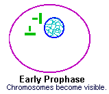
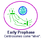
Prophase is the first phase of mitosis but it's named "pro"
which is Greek for "before". That's because the early
cell biologists were not paying close attention and they thought
the phases that followed were the important ones. Don't make the
same mistake they did!
Prophase is composed of a series of very important yet subtle
events.
|
During prophase the chromatin condenses into chromosomes. This
is the distinguishing characteristic of prophase - visible,
disorganized chromosomes.
Prophase is a rather long process and is often divided into early
and late prophase, but the first step is always the appearance
of the chromosomes. At this stage they are made of identical (sister)
chromatids but all you can see are a ball of "threads".
[I think the prophase nucleus looks like it's full of worms! |
|
|
Also early in prophase, the centrosomes come "alive"
with activity. They move to opposite sides of the cell and develop
spindle fibers - long chains of microtubules that will
be used in the next phases to move chromosomes around.
[Spindle fibers look like monofilament fishing line when viewed through a good microscope, but here I've colored them green to remind you that they "grow" from the centrosomes.] During prophase, as the chromosomes continue to condense, the centrosomes move to opposite "poles" dragging their spindles along. |
|
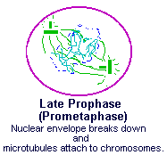
Eventually the cell enters late prophase and, because the next
phase will be metaphase, late prophase is often call prometaphase.
[Recall that "pro" means "before", so this
stage is well named.]
During prometaphase two very important events occur.
Once the microtubules attach to the chromosome they act as a "rope" allowing the centrosome to orchestrate the movements of chromosomes in the next two phases. |
|
This attachment is critical so let's look at it in more
detail.
[It's nearly impossible to see this detail under the microscope
but it is important to understand it.]
|
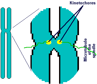
Late in prophase a pair of special structures grow inside of the centromere
region of every chromosome - one on each chromatid.
These structures are called kinetochores.
"Kinetos" is Greek for "movement" and, as
you shall see shortly, that is an excellent name for them.
As a matter of definition - one kinetochore exists on each chromatid so during this phase each chromosome's centromere has two kinetochores. Now the chromosomes are attached to the centrosomes via the spindles! The spindles run from kinetochore (one on each chromatid) to the centrosome (one on each side of the cell). Because there are two kinetochores and two centrosomes, each chromosome is held between the two centrosomes via the spindles. |
|
[If I seem to be beating a dead horse here, please understand that it is important to see how we use the words centromere (on the chromosomes), centrosome (the structures that are now on opposite sides of the cell) and kinetochore (on the chromatid).]
|
Now that the kinetochores of the chromosomes are firmly attached
to the centrosomes, via the spindles of microtubules, we are
ready to understand the critical steps involved in chromosome
movement and division.
The kinetochores have tiny motor proteins that tug on the microtubules (spindle) which are attached to opposite centrosomes. |
|
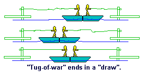
A series of tugging from both kinetochores on their spindles cause each chromosome to move back and forth until this "tug-of-war" ends in a "draw". The chromosome is then positioned midway between the centrosomes. [In point of fact, the kinetochore actually shortens the microtubules so, although there is some pulling involved, it is the shortening of the spindles that contribute to most of the motion.]
|
This cartoon may help you understand.
Imagine each kinetochore is a man standing in a boat (chromatid). Both men hold a rope (spindle fiber made of microtubulin) attached to a nearby dock (the centrosome). The two boats (chromatids) are joined together (at the centromere). As one man (kinetochore) tugs his way, hand over fist, towards his dock, the other man (kinetochore) senses he is drifting away from his dock (centrosome) and makes an effort to drag the boats back to his dock by tugging. |
|
All this back and forth eventually stops once the two kinetochores (men) have positioned the two chromatids that make the one chromosome (their shared boats) midway between the two centrosomes (docks).
|
ALL the chromosomes behave this way.
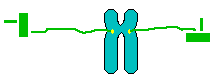
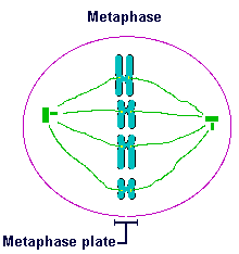
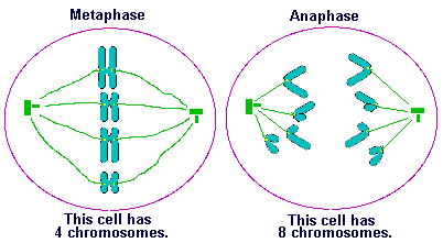
Therefore, all the chromosomes come to line up midway between the two centrosomes. More specifically, when all the chromosomes' centromeres are lying on a plane perpendicular to a line connecting the two centrosomes, the cell has entered the part of mitosis called metaphase. The plane upon which the centromeres are arranged is called the metaphase plate. When you see all the centromeres lined up on the metaphase plate, the cell is in metaphase. The arms of chromosomes don't have kinetochores to guide them and they sometimes flop around. Therefore, the arms and their telomeres might be anywhere but the centromeres must be on the metaphase plate when the cell is in metaphase. |
|
|
Note that in my two dimensional drawing it is easy to see that
the centromeres are all in a line but, technically speaking, metaphase
occurs when they are all in the same plane. This is a technicality
but worth understanding.
Under the microscope they look like they are in a line anyway, because the microscope slide also appears only two dimensional. |
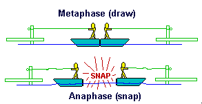
So, during prometaphase (or late prophase) the tug-of-war
is still going on. Once all chromosomes have settled down and
their centromeres are aligned between the centrosomes, the cell
is in metaphase.
Metaphase is well named because "meta" is Greek for
"between".
This is the best time to see chromosomes. Scientists, physicians,
etc. who want to know the numbers, shapes and types of chromosomes
will try to observe cells that are in metaphase.
This phase lasts for about an hour. (Maybe the kinetochores are getting it just right. Or maybe they are getting ready for the big tug that occurs next! )
|
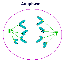
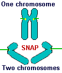
At anaphase the centromere of each chromosome breaks down
the middle (
Let's go back to the cartoon.
It is at anaphase that "true" genetic division occurs. |
|
As each kinetochore drags its chromatid along, the arms of the chromatids are swept back and they seem to form a V-shaped pattern.
This is a matter of definitions BUT it is not a trivial technicality.
It is important because it affects how we count chromosomes.
The trick to counting chromosomes and differentiating them from
chromatids is to keep track of the centromeres. You will recall
that a chromosome has a single centromere and each chromosome
has two kinetochores at the centromere (late in prophase) - one on each chromatid.
|
The moment the chromosome's centromere splits, during anaphase,
each kinetochore acts as the center for the creation of a new
centromere. It's difficult to see in my diagrams, or in a microscope,
but each chromatid is "puckered" inward at the kinetochore
during anaphase and that is the new centromere.
By definition a chromosome has a centromere - it's a one to one correspondence. Therefore, during anaphase, each chromosome becomes two separate chromosomes at that moment of chromatid separation and we now have two chromosomes from each chromosome! |
|
That means, during anaphase and indeed until cytokinesis occurs, the cell has twice as many chromosomes as "normal". But they are all "half-chromosomes" made of just one chromatid.
Read that last section again very slowly and study this diagram to
make sure you understand it.
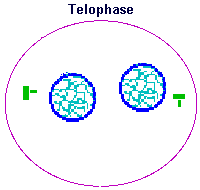
Once the newly created chromosomes reach their poles, anaphase is over and we enter the next and last phase of mitosis - telophase.
"Telos" is Greek for "end". [Good name, huh?  ] ]
Telophase is the point in mitosis when the new nuclei are formed. A nuclear membrane forms around both bundles of chromosomes at each pole. This creates two nuclei. Also, the chromosomes begin to unwind (decondense) until eventually they are nothing more than a dim coloring of chromatin with a few nucleoli (clumps of chromatin). Each nucleus eventually takes on the appearance of an interphase nucleus and once that is done the cell has completed telophase and mitosis. [Phew!] |
|
Mitosis is designed to create two nuclei that are identical in their genetic makeup. They are identical because the sister chromatids at metaphase were identical. Those identical chromatids are the result of the S phase that occurred (long ago, it seems) during interphase but we couldn't see them until prophase or metaphase. At metaphase the chromosomes were carefully lined up along the metaphase plate. Anaphase ensured that one identical chromatid, now a chromosome, made it safely to each pole. Telophase produced the identical nuclei that we now call daughter nuclei because they will be the nuclei of the daughter cells produced from the upcoming cytokinesis.
|
Not only are the daughter nuclei identical to each other but they
are also identical to the mother nucleus from which they came.
The daughter nuclei are clones of each other and clones of their
mother nucleus.
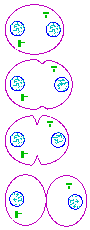
Also, notice that a mother cell must have two centrosomes in order to orchestrate mitosis, but both daughter cells produced, after cytokinesis, have only one centrosome each. When the daughter cells enter late G2, they will duplicate their centrosome and the whole cycle starts again. Take a look at this image on the right, showing cytokinesis, to see what I am talking about. This has been a long and detailed lesson and I have introduced a lot of new and useful concepts. It is important that you understand these ideas and the SAQs will help you with your studies. |
|
and licensed under a Creative Commons Attribution-ShareAlike 4.0 International License.
| Table of Contents | Homepage | How to get a FREE copy of the entire course (hypertextbook) | Frequently Asked Questions |
 ]
]